Sci-Pho
First published in Sanctuary Asia,
Vol. 35
No. 8,
August 2015
Purva Variyar delves into the world of biological photography to showcase natures marvels up-close.
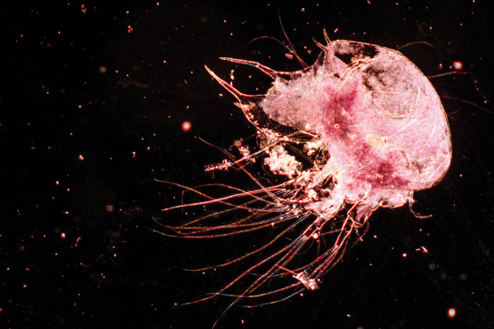
This stunningly beautiful, marine cladoceran crustacean (water flea) looks like its floating amidst a galaxy of stars. The image was made using a microscope, in the dark field, magnified ten times. Photo: Purva Variyar.
For me, the camera has never been about taking pretty photographs. I yearn to capture things that otherwise remain shrouded by our mental and physical limitations of how far we can see and understand what's in front of our eyes.
Science.
I try to express science through my work. Science excites me, and my urge to understand the art of capturing and illustrating ‘science through photographs' led me to pursue an enlightening course, M.Sc. Biological Photography and Imaging from the University of Nottingham.
Look around you and just push yourself a little to wonder about the brilliant and almost perfect mechanics behind everything that you see.
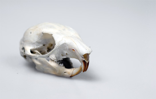
A skull can reveal so much about an animal, for that matter, an entire species. This skull of an unidentified species of a squirrel made for an interesting subject. Photographing it in a studio replete with lights and background gave it a professional touch. Photo: Purva Variyar.
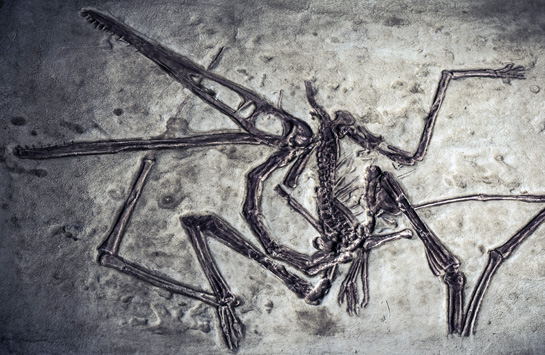
Fossils! I love them. The mystery and stories they hold have me under their ‘sorcery spell. This fossil replica of the flying reptile Pterodactylus Pterodactylus antiquus was simply too irresistible to not photograph. Photo: Purva Variyar.
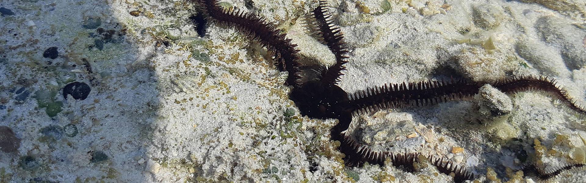
If you dig deep, there is a lot going on under the radar which is far more interesting than what is obvious. When you see a snail, do you stop to think how it came to be? How and what went into this life form to allow it to emerge from practically nothing to quite something. To learn about wildlife and natural history, one need not always spend time in the forest. The incredible array of natural history specimens in laboratories and museums will stun you. Try the Bombay Natural History Society for example. You wouldnt believe the things you can see and learn here, the exciting facts and discoveries awaiting your encounter.
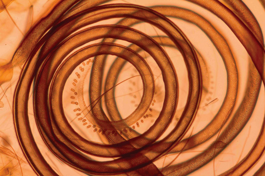
Everything under the microscope looks more magical than it already is, and brings out details hitherto unimaginable. So much escapes our eyes. This beautiful, coiled thing is the proboscis of a unidentified butterfly species, taken in phase contrast and magnified five times. Photo: Purva Variyar.
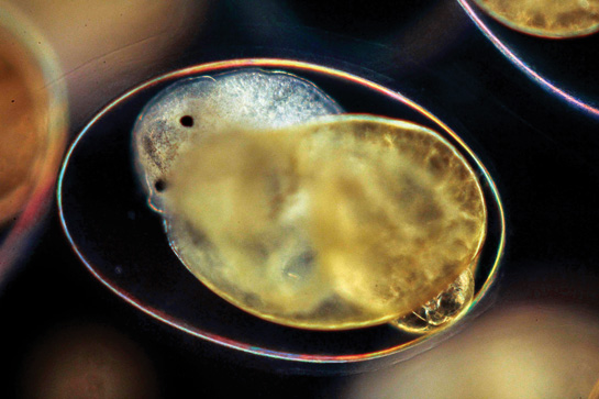
I was left open mouthed when I first saw this beauty under the microscope. Normally, to study a specimen or cells under a microscope, they have to be in a dead state. But, thanks to the transparent egg capsule of the Lymnaea stagnalis, a pondwater species of snail, I got to see live snail embryos still in the eggs, with their tiny hearts beating! Photo: Purva Variyar.
Purva Variyar is a conservationist, writer, and editor. Her true passion lies in science communication. She currently works with the Wildlife Conservation Trust. Purva has previously worked as a Senior Editor and Science Communicator with Sanctuary Asia magazine. She has also worked on human-snake conflict mitigation and radio-telemetry of Russell’s vipers under The Gerry Martin Project. To see more of Purva's images visit her Facebook page Sci-Pho.
First appeared in: Sanctuary Asia, Vol. 35 No. 8, August 2015.